Difference between revisions of "Hyper Rayleigh Scattering"
Cmditradmin (talk | contribs) m (→Technique) |
Cmditradmin (talk | contribs) m |
||
| Line 13: | Line 13: | ||
See Firestone 2004 <ref>K. A. Firestone, P. Reid, R. Lawson, S. H. Jang, and L. R. Dalton, “Advances in Organic Electro-Optic Materials and Processing,” Inorg. Chem. Acta, 357, 3957-66 (2004)</ref>. | See Firestone 2004 <ref>K. A. Firestone, P. Reid, R. Lawson, S. H. Jang, and L. R. Dalton, “Advances in Organic Electro-Optic Materials and Processing,” Inorg. Chem. Acta, 357, 3957-66 (2004)</ref>. | ||
There are a number of ways the HRS | There are a number of ways the HRS setupcharacterization is used in non-linear optics research. | ||
The dielectric properties of the solvent influence the β (solvatochromatism). | The dielectric properties of the solvent influence the β (solvatochromatism). One area of research involves predicting and solvent effects so as to better understand the performance of molecules when densely packed in materials. Attempts have been made to calibrate HRS measurements with EFISH measurements of beta though they are based on different tensor elements. | ||
See Wikipedia on [http://en.wikipedia.org/wiki/Rayleigh_scattering Rayleigh Scattering] | See Wikipedia on [http://en.wikipedia.org/wiki/Rayleigh_scattering Rayleigh Scattering] | ||
Revision as of 15:46, 12 October 2009
Hyper Rayleigh Scattering (aka Harmonic Light Scattering or HRS) is one method for measuring the first hyperpolarizabilityβ. Another method is electric field induced second harmonic generation (EFISH)
Overview
In HRS A dilute sample of a test chromophore is prepared in a solvent. An incident laser generates a second harmonic signal, specifically the frequency double signal from excitation of the sample molecules. This can be related to the beta of the sample using this formula:
- <math>\frac {I_{sample}} {I_{solvent}} = \frac {N_{sample} \langle \beta^2 _{sample} \rangle + N_{solvent} \langle \beta^2_{solvent}\rangle} {N_{solvent} \langle \beta^2_{solvent}\rangle}\,\!</math>
See Firestone 2004 [1].
There are a number of ways the HRS setupcharacterization is used in non-linear optics research.
The dielectric properties of the solvent influence the β (solvatochromatism). One area of research involves predicting and solvent effects so as to better understand the performance of molecules when densely packed in materials. Attempts have been made to calibrate HRS measurements with EFISH measurements of beta though they are based on different tensor elements.
See Wikipedia on Rayleigh Scattering
See also Density Functional Theory
See Wikipedia on Raman Scattering
Technique
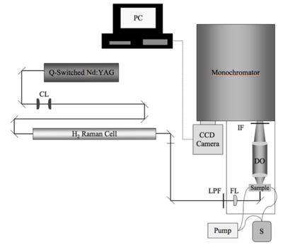
The HRS measurement of β is a specialized device custom-built on an optics table. The components and configuration can change significant from site to site. It is important to understand the significance of each portion of the setup. One possible HRS experimental setup is described here.
- Q-switched Nd:YAG laser operating at 1064 nm with a repetition rate of 30 Hz is directed along a double-back path in order to make it easier to adjust the optics along the path.
- The beam is directed into a 1-m H2 Raman cell which produces 1907 nm (the first stokes line - near infrared) radiation. The desired wavelength is isolated Pellin-Broca prism, dichroic mirrors, and a Corning long-pass emission filter resulting in an incident power of 400mW.
- The exciting beam is directed into a 10mm flow cell through which fresh sample is continually pumped so as to avoid the effects of photo degradation of the sample.
- Light scattered at 90 degrees from the excitation beam path is focused into a monochromatic containing a 1200 g/mm ruled diffraction grating and 950nm interference filter. This isolates radiation emitted at the second harmonic (2 omega) of the excitatory beam ( lambda/2= 1907 /2=950)
- Finally the specific desired wavelength was measured with a CCD camera with long exposures of 240-540 seconds.
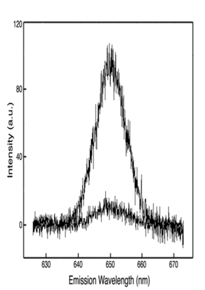
- The HRS spectra is processed to remove low intensity background two-photon fluorescence which can be fitted to a fourth order polynomial and then subtracted.
- The final HRS spectra is fitted to a Gaussian functional form.
- At this point a number of mathematical manipulations and comparisons can be done, for example comparing the sample HRS to a known reference sample with known beta, or internal referencing of the solvent contribution of beta.
Video to come
Significance
References
- ↑ K. A. Firestone, P. Reid, R. Lawson, S. H. Jang, and L. R. Dalton, “Advances in Organic Electro-Optic Materials and Processing,” Inorg. Chem. Acta, 357, 3957-66 (2004)

